

You show up for your eye exam. The tech asks: "Do you want the dilation drops, or would you prefer the retinal photo?" People often choose retinal photos to avoid the glare and blur of dilating drops. But these are not the same test, and the wrong answer for your situation could mean something gets missed.
Here's a clear breakdown of what each one does, what each one catches, and how to know which one — or both — you actually need.
What Is a Dilated Eye Exam?
A dilated eye exam uses medicated eye drops — typically tropicamide or phenylephrine — to temporarily relax the muscles that control your iris. This pharmacological intervention typically requires 15 to 30 minutes to achieve full dilation, after which your eye doctor uses a bright light and specialized lenses to examine the retina, optic nerve, and blood vessels at the back of the eye.
The side effects are real and last a while. Light sensitivity and blurry vision can last the rest of the day, and you might need to wear sunglasses indoors or avoid driving after the appointment.
What you get in exchange: a comprehensive, direct, magnified view of your retinal structures — the closest thing to putting a doctor's eyes inside your eye.

What Is Retinal Photography?
Retinal photography (often called fundus photography or Optomap imaging) uses a specialized digital camera to capture high-resolution images of the back of your eye — no drops required. The most widely recognized system is ultra-widefield imaging technology, such as Optomap, which can document up to 82% of the retina in a single capture lasting less than half a second.
The retina is the only area of the human body where doctors can examine blood vessels without cutting open the body, which makes retinal imaging useful for detecting signs of systemic conditions like diabetes, hypertension, and high cholesterol — not just eye disease.
The procedure is fast and painless. No blurry vision afterward, no light sensitivity, no need for a ride home. It also produces a permanent digital record that your doctor can compare year over year to track any changes.
Dilated Exam vs Retinal Photo: Side-by-Side
| Feature | Dilated Eye Exam | Retinal Photography |
|---|---|---|
| Eye drops required | Yes | No |
| Wait time | 15–30 min for dilation | Under 1 minute |
| Recovery time | 4–8 hours (blurred vision, light sensitivity) | None |
| Retinal coverage | Up to 240 degrees | Up to 200–270 degrees depending on system |
| Optic nerve magnification | High — clinical gold standard | Lower magnification |
| Stored digital record | No | Yes |
| Additional cost | Usually covered by exam fee | Often not covered by vision insurance |
| Best for | High-risk patients, complete clinical evaluation | Baseline screening, monitoring, convenience |

What Each One Can (and Can't) Detect
Both methods look at the same real estate — the retina, optic nerve, and blood vessels — but they have different strengths.
Dilated Exam
Dilation offers the most thorough assessment of eye and overall health and is particularly essential for high-risk patients. It provides a magnified, three-dimensional view of the optic nerve, which matters when screening for glaucoma. It also gives your doctor a direct view of the peripheral retina, where retinal tears and early detachments often begin.
A dilated eye exam provides a more magnified view of the optic nerve. Because Optomap imaging is focused on the retina, it provides less magnification of the optic nerve, which means some signs of glaucoma can be missed with retinal photography alone.
Retinal Photography
Research demonstrates that retinal imaging can detect subtle changes that might be missed during routine examinations, particularly microaneurysms, small hemorrhages, and early drusen formation associated with macular degeneration.
The optomap images can be stored and compared for changes over time, making it especially useful for monitoring known conditions or establishing a baseline in younger patients.
One important distinction: retinal cameras can magnify images significantly, which means tiny findings — pinpoint hemorrhages, microaneurysms, and Hollenhorst plaques — are often more easily spotted via retinal photography than through a dilated fundus exam. Photos also create a permanent record your doctor can show you directly, and compare against your next visit to track whether anything has changed.
Where dilation has the clear edge is in viewing structures in front of the retina. If your doctor needs to assess cataracts or other anterior structures, dilation is the better tool — and that's precisely why some patients end up doing both in the same visit.
Who Specifically Needs Dilation
Retinal photography works well as a screening tool for healthy, low-risk adults. But certain patients should not skip dilation. Patients with high prescriptions, children aged 10 and under, patients aged 60 and over, patients with retinal issues, and patients with certain disease states including but not limited to diabetes will all require dilation in addition to retinal imaging.
If you have high myopia (above approximately -5.00 diopters), the stakes are elevated. Flashing lights or new floaters warrant an urgent dilated exam to rule out retinal tears or detachment — dilation gives the widest peripheral view, which is where those problems tend to show up first. For glaucoma monitoring, retinal photos of the optic nerve are a valuable addition, since a documented baseline image makes it far easier to detect subtle changes at the next visit.
For diabetics, retinal photography is particularly strong at picking up diabetic retinopathy — the camera can catch microaneurysms and small hemorrhages with a level of detail that makes it a reliable screening tool. That said, dilation still adds value: better peripheral retinal coverage and the ability to assess the lens for cataracts, which diabetics are at higher risk for. For high-risk patients in general, the most thorough approach is both — photos for documentation and fine detail, dilation for peripheral access and structures in front of the retina.
Do Contact Lenses Affect Pupil Dilation?
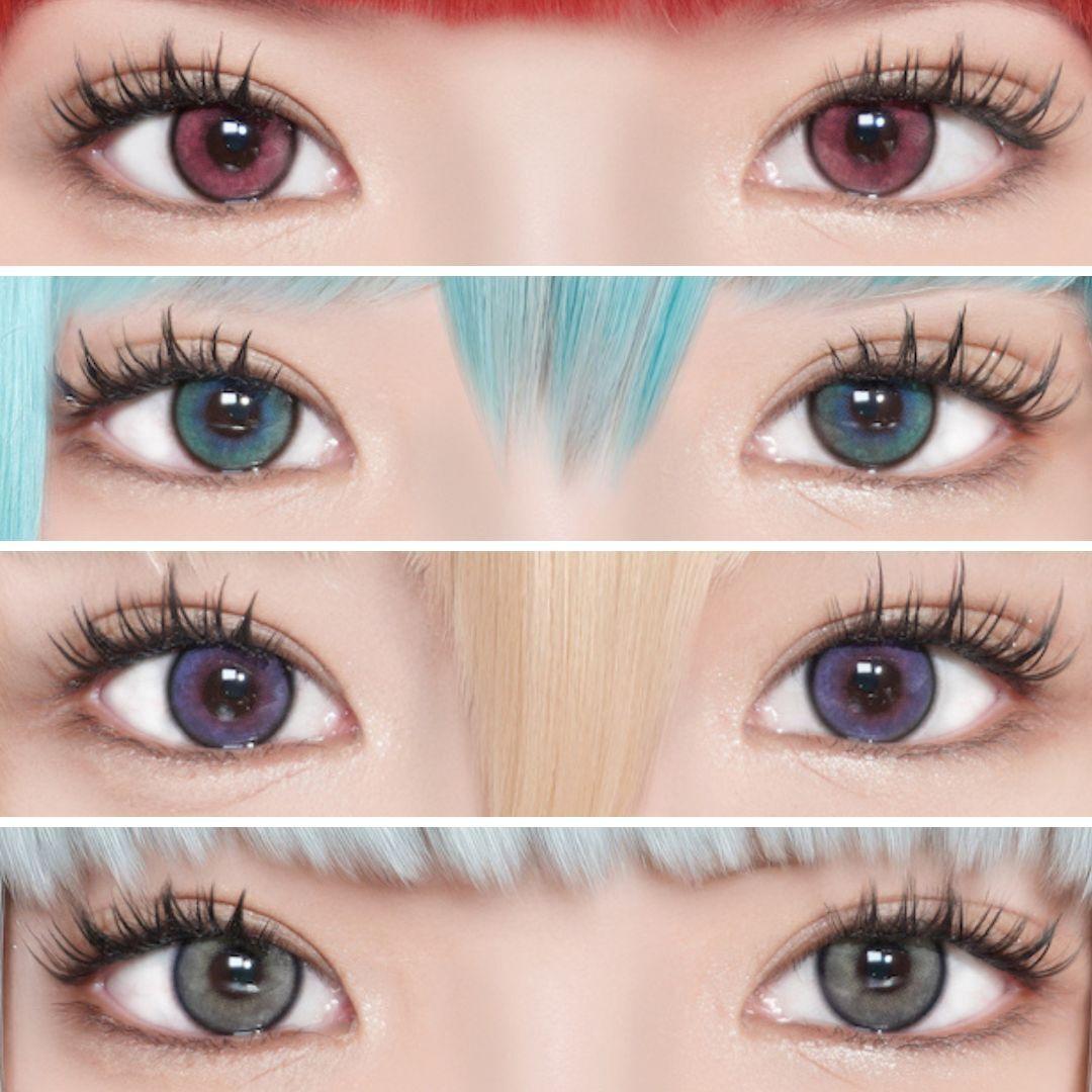
Short answer: soft contact lenses should be removed before dilation drops are applied, but they don't prevent dilation from working. Your doctor will ask you to take them out before administering the drops. This is standard procedure whether you wear clear prescription contacts or colored contacts.
After the exam, wait until your vision has fully cleared before putting lenses back in — the dilating drops temporarily affect your ability to focus, and inserting contacts before that resolves can be uncomfortable and creates a higher risk of improper placement.
If you wear rigid gas-permeable (RGP) lenses, your doctor may have you remove them ahead of the exam for an extended period, as RGP wear can temporarily alter corneal shape and affect certain measurements.

Can Retinal Photos Replace Dilation?
For most healthy adults under 40 with no systemic disease and no family history of eye conditions, retinal photography provides a solid annual screening baseline. Retinal imaging provides excellent baseline documentation and patient engagement opportunities, particularly for younger, healthy adults. Dilation remains essential for complete assessment, especially in patients with symptoms, risk factors, or complex medical histories.
Optomap and dilation are not rivals — they're teammates. Optomap captures a wide retinal image quickly and comfortably, but some areas require a closer look, and dilation provides that more detailed view.
The most accurate answer is: it depends on your risk profile. If you're young, healthy, no diabetes, no family history of glaucoma or macular degeneration, and you have a low-to-moderate prescription, retinal photography is a reasonable annual option. If any of those conditions apply, insist on dilation — or ideally, both.
Frequently Asked Questions
Is a dilated eye exam required to get a contact lens prescription?
Not always, but it depends on your eye doctor's protocol and your health history. A comprehensive contact lens exam includes a refraction and fit assessment, and may or may not include dilation. Patients with higher prescriptions or health risk factors are more likely to require it.
How long does pupil dilation last?
Typically 4 to 8 hours, though it varies by individual and the specific drops used. Light sensitivity and difficulty focusing on close objects are the main effects during this period.
Does retinal photography hurt?
No. It involves a brief flash of light and takes under a minute. There are no drops and no recovery period.
Can I drive after a dilated eye exam?
You should avoid driving until your vision has fully returned to normal. Most patients arrange a ride or wait several hours before driving.
Why does my eye doctor charge extra for retinal photos?
The cost of retinal photography is often not covered by vision insurance, so it typically appears as an add-on fee. Dilation is usually included in a comprehensive exam co-pay.
If I wear colored contacts, do I need to remove them for a retinal exam?
Yes. Whether you're getting dilation or retinal photography, contact lenses should be removed before either procedure. Both require a clear, unobstructed view of the eye.
Keep Your Prescription Current
Whether you need dilation or a retinal photo, keeping your prescription current is step one. Browse EyeCandys' range of FDA-cleared prescription contact lenses and colored contacts — comfort and clarity.