

Most people assume color blindness means seeing the world in black and white. It doesn't. The vast majority of people with color vision deficiency (CVD) see color — just differently. Some can't distinguish red from green. Others confuse blue and yellow. A very small number genuinely see almost no color at all.
Color vision deficiency affects roughly 1 in 12 males and 1 in 200 females worldwide. That's approximately 300 million people. Yet most go years — sometimes their entire lives — without a formal diagnosis, because the vision they've always had is the only vision they know.
This guide breaks down every major type of color blindness: what's happening in the eye, what the world actually looks like, how common each type is, and what (if anything) can be done about it.
What Is Color Blindness, Really?
The term "color blindness" is a bit of a misnomer. The clinical term — color vision deficiency — is more accurate for most cases. Total color blindness (seeing only shades of grey) is extremely rare.
Color vision works through photoreceptor cells in the retina called cones. Humans have three types:
- L-cones — sensitive to long wavelengths (red light, ~560 nm)
- M-cones — sensitive to medium wavelengths (green light, ~530 nm)
- S-cones — sensitive to short wavelengths (blue light, ~420 nm)
When all three function normally, the brain combines their signals to distinguish approximately one million shades of color. Color vision deficiency occurs when one or more cone types is absent, reduced in sensitivity, or shifted in its spectral response — creating overlap or gaps in color perception.
Why does it affect more men? The genes encoding L-cones and M-cones sit on the X chromosome. Males have one X chromosome (XY), so a single defective copy causes red-green CVD. Females (XX) need both X chromosomes to carry the defect — which is why red-green color blindness affects around 8% of males but only about 0.5% of females. Blue-yellow CVD involves a gene on chromosome 7 (an autosome), so it affects both sexes equally.

The Two Main Categories
Before getting into specific types, it helps to understand the two severity tiers:
Anomalous Trichromacy
All three cone types are present, but one is shifted in sensitivity. Colors are still perceived, just with reduced discrimination. This is the milder, more common form.
Dichromacy
One cone type is entirely absent. Certain colors become genuinely indistinguishable from one another, not just difficult to tell apart.
Monochromacy
Two or all three cone types are absent or nonfunctional. Extremely rare. This is where true "colorblindness" actually lives.
Red-Green Color Blindness (The Most Common Type)
Red-green color blindness accounts for roughly 95% of all color vision deficiencies. It comes in four subtypes — two affecting red perception (protan defects) and two affecting green (deutan defects).
Protanomaly — Weak Red Cones
Protanomaly is an anomalous trichromacy condition where L-cones are present but shifted toward green sensitivity. Red appears less bright and less saturated than normal — often looking more like a muted orange or brownish tone. Colors containing red (orange, purple, pink) also appear shifted. Protanomaly affects approximately 1% of males.
Protanopia — Missing Red Cones
Protanopia is the dichromatic version: L-cones are absent entirely. Red is essentially invisible as a distinct color. Most reds appear as dark yellow or brown; greens look similar to yellows. The entire red-to-green range collapses into a narrower spectrum of blues and golds. Protanopia affects roughly 1% of males.
Deuteranomaly — Weak Green Cones
Deuteranomaly is the most common subtype of color blindness overall, affecting approximately 5% of males. M-cones are present but shifted toward red sensitivity. Greens, yellows, oranges, and reds can all appear similar — particularly in low-light conditions. Blues and purples may also be hard to distinguish.
Deuteranopia — Missing Green Cones
With deuteranopia, M-cones are absent. The result is similar to protanopia in terms of the red-green confusion, though reds appear slightly less dimmed than in protanopia because L-cones are still functional. Deuteranopia affects approximately 1% of males.

Blue-Yellow Color Blindness (Tritan Defects)
Blue-yellow color blindness is far rarer than red-green CVD, affecting fewer than 1 in 10,000 people. Critically, because the gene involved (OPN1SW) sits on chromosome 7 rather than the X chromosome, it affects males and females equally. It's also more frequently acquired than inherited — triggers include aging, certain medications, glaucoma, and retinal disease.
Tritanomaly — Weak Blue Cones
Tritanomaly involves reduced S-cone sensitivity. Blues appear shifted toward green; yellows may look indistinguishable from light pinks or greys. The defect is mild enough that many people are unaware they have it. Prevalence is estimated below 0.01%.
Tritanopia — Missing Blue Cones
Tritanopia is the dichromatic form. S-cones are absent. Blue and green become difficult or impossible to distinguish; dark blue and black can look identical. Yellows shift toward pinks or light greys. Like tritanomaly, prevalence sits below 0.01% of the population.

Complete Color Blindness (Monochromacy)
Achromatopsia
Achromatopsia is the condition most people picture when they think of color blindness — seeing the world in near-greyscale. It results from a complete absence of functioning cone cells, leaving vision reliant entirely on rod photoreceptors. Rods handle low-light vision and are not sensitive to color.
Beyond the absence of color, achromatopsia typically comes with reduced visual acuity, extreme light sensitivity (photophobia), and involuntary eye movement (nystagmus). It's an autosomal recessive disorder, meaning both copies of the defective gene must be inherited. Prevalence is approximately 1 in 30,000 people.
Blue Cone Monochromacy
Blue cone monochromacy is an X-linked condition in which both L- and M-cones fail to function normally, leaving only S-cones (blue-sensitive) active. Like achromatopsia, it results in very poor color discrimination, reduced acuity, nystagmus, and photophobia — though some residual blue-range color perception remains. It is rarer than achromatopsia.
How Is Color Blindness Diagnosed?
A standard eye chart test will not detect CVD. Specific color vision tests are needed:
| Test | What It Detects | Limitations |
|---|---|---|
| Ishihara test | Red-green CVD screening | Cannot detect blue-yellow deficiency or distinguish protan from deutan subtypes |
| Farnsworth-Munsell 100 Hue | All CVD types; measures severity | More time-intensive; requires clinical setting |
| Anomaloscope | Protan and deutan classification (gold standard) | Mainly used in research and specialist settings |
If you suspect you have a color vision deficiency, an optometrist or ophthalmologist can administer these tests and confirm the type and severity.

Can Color Blindness Be Corrected?
There is currently no medical cure for inherited color vision deficiency. Research into gene therapy is ongoing, but no approved treatment exists.
Filter glasses — brands like EnChroma use optical notch filters that selectively block wavelengths in the 530–560 nm range where L- and M-cone sensitivities overlap. The effect is increased contrast between red and green signals for people with anomalous trichromacy (protanomaly or deuteranomaly). Studies show improvements in color discrimination for some users, particularly those with mild-to-moderate red-green CVD. They do not restore normal color vision and have little to no effect on dichromats (protanopia, deuteranopia) or tritan defects.
Tinted contact lenses — research-stage work has explored incorporating wavelength-filtering dyes into contact lens materials to achieve similar effects to filter glasses, with the advantage of convenience and cosmetic subtlety. Studies have shown some improvement in color discrimination in lab settings. However, as of now, no FDA-cleared color-correcting contact lens is commercially available for CVD. Standard colored cosmetic contact lenses do not correct color blindness.
Assistive apps and tools — smartphone apps like Colorblind Avenger and Be My Eyes use camera input to identify and name colors, helping users navigate color-dependent tasks in daily life.
Living With Color Vision Deficiency
For most people, color vision deficiency is a minor functional difference rather than a disability. The brain adapts — people learn to use contextual cues (brightness, position, shape) to interpret color information they can't directly perceive. A red traffic light is always on top; a ripe banana has a shape you recognize before you register its color.
Daily challenges do exist: reading color-coded charts, selecting produce, choosing clothing that matches, and navigating certain digital interfaces where color alone conveys information. Some professions — commercial aviation, certain military roles, electrical wiring, graphic design — have color vision requirements that CVD can affect.
The most useful first step for anyone who suspects a color vision deficiency is a proper clinical test. Knowing your specific type and severity opens up better options for management, workplace accommodation, and everyday adaptation.
Frequently Asked Questions
What is the most common type of color blindness?
Deuteranomaly — a form of red-green color blindness caused by shifted green cone sensitivity — is the most common subtype, affecting approximately 5% of males.
Is color blindness more common in men or women?
Yes, significantly. Red-green color blindness affects around 8% of males and only 0.5% of females, because the responsible genes are located on the X chromosome. Males have one X chromosome, so a single defective copy is sufficient to cause the condition.
Can you get contact lenses for color blindness?
Specialty tinted contact lenses exist (such as the X-Chrom lens) and have been used in clinical settings to alter color perception for some patients with red-green CVD. Research into dye-based filtering contacts is ongoing. However, no commercially available, FDA-cleared contact lens currently corrects color vision deficiency. Standard cosmetic colored contacts do not have this effect.
Is color blindness hereditary?
In most cases, yes, but not always. Red-green color blindness is an X-linked recessive condition inherited through the mother. Blue-yellow color blindness is autosomal dominant and can be inherited from either parent. Achromatopsia is autosomal recessive. Some forms of CVD can also be caused by acquired eye disease (e.g. cataracts, glaucoma and macular degeneration), medication, or aging.
What's the difference between color blindness and color vision deficiency?
They refer to the same group of conditions, but "color vision deficiency" (CVD) is the more clinically precise term. "Color blindness" implies a total absence of color vision, which is only true in rare cases like achromatopsia. Most people with CVD perceive color — just with reduced ability to distinguish certain hues.


Color Contacts for Every Look
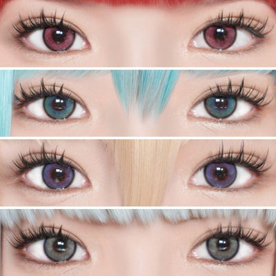
Whether you're after a subtle enhancement or a bold transformation, EyeCandys carries hundreds of colored contact lens styles — from natural-looking tints to vivid statement lenses. FDA-cleared.