


Some people may think an eye appointment is about getting a new prescription. It's not — or at least, that's not all it is. A dilated eye exam is one of the few non-invasive windows into your body's internal health, and what your eye doctor can find there goes far beyond whether you need new glasses.
Your eyes are the only place in the body where blood vessels, nerve tissue, and central nervous system tissue can be directly visualized without invasive procedures. That makes the back of your eye — the retina, optic nerve, and surrounding vasculature — an extraordinarily useful diagnostic surface. Conditions that are quietly developing elsewhere in your body often leave their earliest fingerprints here, sometimes years before symptoms show up anywhere else.
Here's what a dilated eye exam can actually catch.
What Happens During Eye Dilation
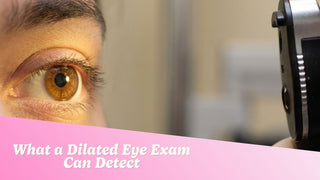
Your eye doctor uses prescription drops to temporarily widen your pupils. Dilating eyedrops allow the doctor to typically see much more of the retina than without dilating. Many practices also offer special cameras that can photograph most of the retina without drops — however, these cameras may miss parts of the peripheral retina and don't allow visualization of other structures like a cataract.
The drops take about 15–20 minutes to work. Your pupils will remain dilated for 4 to 6 hours after the exam, causing light sensitivity and blurred near vision. Plan for it — bring sunglasses and, if possible, a driver.
Eye-Specific Conditions It Catches
These are the conditions commonly associated with eye exams, and they're serious enough on their own.
Glaucoma
Glaucoma often has no early warning signs, making dilated exams essential for detection. The optic nerve — more visible through a dilated pupil — shows characteristic changes when intraocular pressure has been building. Caught early, the progression can be slowed significantly. Missed, it causes irreversible vision loss.
Diabetic Retinopathy
The blood vessels in the retina can leak blood, which your optometrist can see during a dilated examination. Diabetic retinopathy is the leading cause of blindness in working-age adults, and its early signs appear in the eye before patients have any noticeable vision symptoms.
Macular Degeneration
The macula controls central, detail vision — reading, driving, face recognition. Macular degeneration can cause significant vision loss, but when caught early through dilated exams, treatments can slow its progression.
Retinal Tears and Detachment
The peripheral retina — the zone most likely to develop tears — is only fully accessible with a dilated pupil. A tear caught before it becomes a detachment is treated with a laser procedure. A detachment is a surgical emergency with a real risk of permanent vision loss.

The Systemic Conditions: What Most People Don't Know
This is where dilated eye exams earn their status as a public health tool. According to the American Optometric Association, a dilated eye exam can detect more than 270 diseases and abnormalities.
Diabetes and Pre-Diabetes
Signs of diabetic retinopathy — such as tiny blood vessels in the retina leaking yellow fluid — may appear in eye tissue even before a person has been diagnosed with diabetes. In 2017, optometrists diagnosed more than 400,000 patients with diabetic retinopathy in people who hadn't yet been diagnosed with the condition. A comprehensive eye exam was their first signal that something was wrong.
High Blood Pressure (Hypertension)
A dilated eye exam can reveal uncontrolled high blood pressure, which increases your risk of heart attack and stroke. During an exam, the very small blood vessels look narrowed in the retina — instead of looking red, they appear slightly pink or silvery, and much narrower than expected. Signs include "AV nicking," where a stiffened artery compresses a vein where they cross, generalized narrowing of the retinal arteries, and retinal hemorrhages. Since hypertension is typically symptomless until it causes a crisis, the eye exam can be a literal lifesaver.
High Cholesterol
A yellow or blue ring around the cornea may be a sign of high cholesterol. Deposits can also appear in the blood vessels of the retina. Tiny cholesterol emboli (Hollenhorst plaques) can sometimes be seen lodged within retinal arteries, signaling a significant risk of stroke.
Stroke Risk
An ophthalmologist or optometrist can spot plaque deposits or blood clots in the arteries of the eye. These point to an increased risk of stroke if the deposits reach the brain. A loss of peripheral vision may also be a warning of brain damage caused by a previous stroke.
Brain Tumors and Elevated Intracranial Pressure
Increased pressure inside the skull can cause visible swelling in the optic nerve — a condition called papilledema — which is detectable during a dilated eye exam. Causes include brain tumors, aneurysms, and idiopathic intracranial hypertension (IIH), a condition that most commonly affects young women with obesity where pressure builds without a clear underlying cause. This is one of the more striking capabilities of the exam: a non-invasive look at a direct extension of your brain.
Thyroid Disease
Graves' disease, the most common eye disease associated with a dysfunctional thyroid, is typified by protruding eyeballs or retracting eyelids, occurring when there is too much or too little thyroid hormone. These are often among the first visible signs of a thyroid disorder.
Autoimmune Conditions
The systemic conditions actively screened for during a comprehensive eye exam include autoimmune conditions such as rheumatoid arthritis, lupus, and multiple sclerosis. In rheumatoid arthritis, the most common ocular manifestation is keratoconjunctivitis sicca (dry eye). In multiple sclerosis, it is optic neuritis — inflammation of the optic nerve that can cause sudden vision loss. These signs sometimes appear before a formal diagnosis has been made elsewhere.
Cancer
Cancer can potentially show up in and around the eyes. The most common skin cancers — basal cell, squamous cell, or melanoma — may be present on the outer surfaces of the eye or the eyelids. Lymphoma or leukemia can cause changes in the interior of the eye. Breast cancer and other bodily cancers can metastasize and spread to the eye and its structures.

How Often You Actually Need an Eye Exam
Frequency depends on your age and risk profile:
-
Ages 18–40: every 2 years, or as recommended by your optometrist
-
Ages 40+: every year, as risks for eye diseases increase
-
Diabetes, high blood pressure, or family history of eye disease: annually or as directed by your eye doctor
If you wear contact lenses, this applies to you too. Your annual contact lens fitting appointment is not a substitute for a comprehensive dilated exam — they're different things, and your eye care provider should be doing both.

After the Exam: What to Expect
Plan your day around it. Light sensitivity and blurred near vision are normal for several hours. Wearing sunglasses and avoiding screens can help ease discomfort. Whether you can drive depends on how much dilation affects your vision personally — when in doubt, arrange a ride.
If you wear contacts, bring your glasses. You'll want to remove your lenses before the drops go in, and your vision will be too blurry for safe lens handling immediately after.

Frequently Asked Questions
Is a dilated eye exam the same as a regular eye exam?
No. A vision test checks your prescription. A dilated exam uses drops to widen your pupils and typically gives your eye doctor a more detailed view of the retina and inside of the eyes versus without dilation. The two exams serve different purposes and are ideally done together.
Can a dilated eye exam detect diabetes?
Yes. Signs of diabetic retinopathy — tiny leaking blood vessels in the retina — can appear before a person has even been diagnosed with diabetes. Many people have received a diabetes referral from their eye doctor before their primary care physician caught it.
How long does dilation last?
Typically 4 to 6 hours. Expect light sensitivity and blurred near vision during that window.
Can I drive after a dilated eye exam?
It depends on the individual. Many people are fine to drive; others find the light sensitivity too disruptive. If it's your first time, arrange a ride or plan to wait it out before getting behind the wheel.
Can I wear contacts to a dilated eye exam?
Bring them, but you'll need to remove your lenses before the drops go in. Bring your glasses for after the appointment.
How many conditions can a dilated eye exam detect?
According to the American Optometric Association, more than 270 diseases and abnormalities can be identified through a dilated eye exam.
Do I need a dilated exam if my vision seems fine?
Yes. Most of the systemic conditions described in this article — diabetes, hypertension, glaucoma, early macular degeneration — produce no noticeable symptoms in their early stages. 'Seems fine' isn't the same as 'is fine.'
Keep Your Eyes Healthy. Keep Your Prescription Current.
Regular eye care and the right contact lenses go hand in hand. Browse EyeCandys' full range of FDA-cleared prescription and plano contact lenses — comfort and clarity.